

Contents
I. Review-X1 System
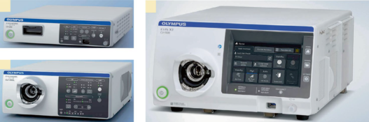
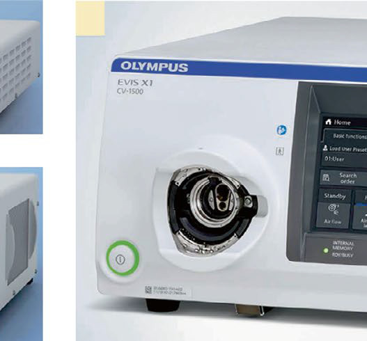
- EVIS X1 Endoscopy System
- Texture and Color Enhancement Imaging (TXI)
- Naoto Tamai and Kazuki Sumiyama
- Red Dichromatic Imaging (RDI)
- Kurato Miyazaki and Motohiko Kato
- Artificial Intelligence (AI) in Colonoscopy
- Masashi Misawa and Shin-ei Kudo
II. Classification
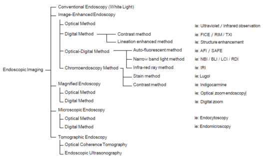
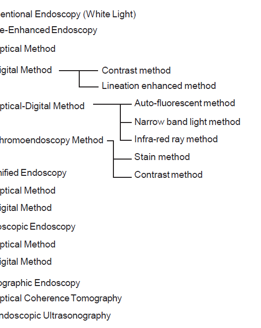
- Classification of Endoscopic Imaging
- Hisao Tajiri
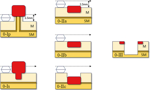
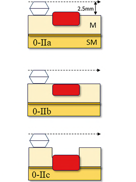
- Paris Classification
- Mineo Iwatate and Santa Hattori
- Esophagus: The Japan Esophageal Society (JES) Classification
- Kenichi Goda and Tsuneo Oyama
- Esophagus: The BING Classification
- Jin Lin Tan and Rajvinder Singh
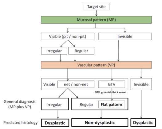
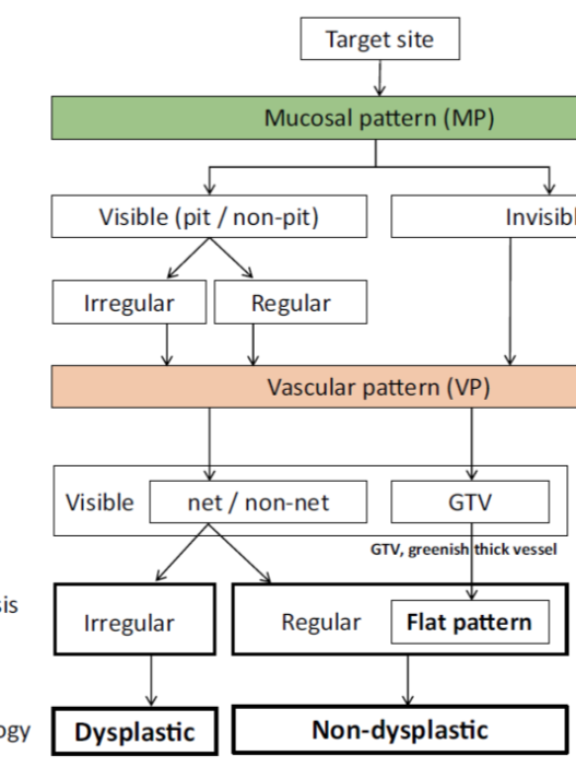
- Stomach: VS Classification/MESDA-G
- Kenshi Yao
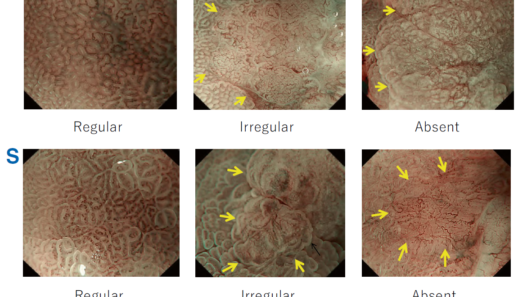
- Colon: The Japan NBI Expert Team (JNET) Classification
- Daizen Hirata and Yutaka Saito
III. High-Quality Photography
- How to Take High-Quality Images (H&N)
- How to Take High-Quality Images (UGI)
- How to Take High-Quality Images (LGI)
IV. Head and Neck
- Pharyngeal Lymphoid Follicle
- Pharyngeal Papilloma
- Pharyngeal Superficial Cancer (0-I)
- Pharyngeal Superficial Cancer (0-IIa)
- Pharyngeal Superficial Cancer (0-IIb)
V. Esophagus (Non-Barrett)
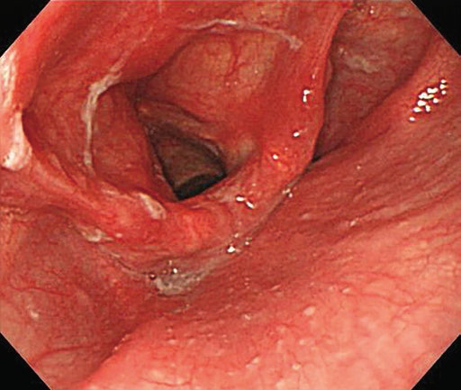
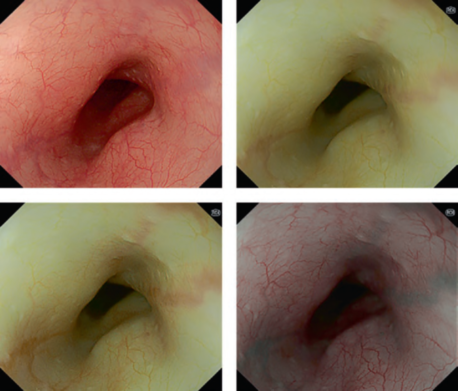
- Reflux Esophagitis
- Esophageal Papilloma
- Multiple Lugol-Voiding Lesions
- High- Grade Squamous Dysplasia of the Esophagus
- Esophageal Squamous Cell Carcinoma (0-I)
- Esophageal Squamous Cell Carcinoma (0-IIa)
- Esophageal Squamous Cell Carcinoma (0-IIc)
VI Barrett’s Esophagus
- Long- and Short-Segment Barrett’s Esophagus
- Barrett’s Esophagus with Low-Grade Dysplasia
- Barrett’s Esophagus with High-Grade Dysplasia
- Barrett’s Dysplasia and Focal Adenocarcinoma
- Barrett’s Adenocarcinoma (0-IIa)
- Barrett’s Adenocarcinoma (0-IIc)
VII Stomach
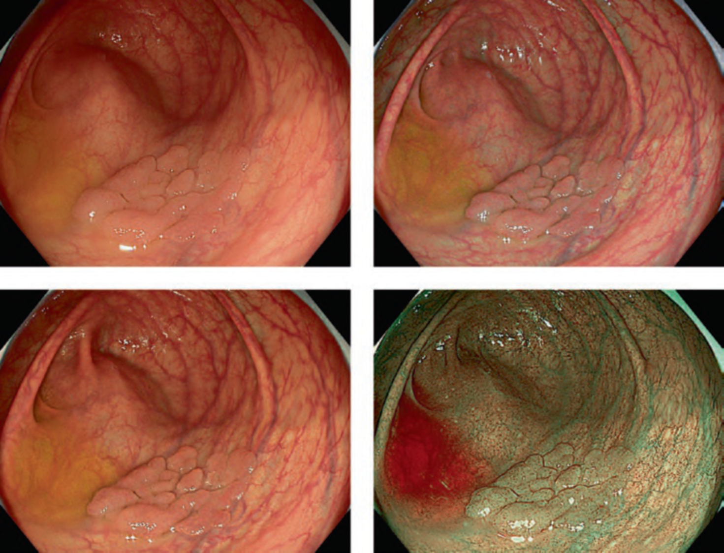
- Gastric Intestinal Metaplasia
- Gastric Hyperplastic Polyp with Differentiated Intramucosal
- Adenocarcinoma
- Gastric Adenoma
- Early Gastric Cancer (0-I, Differentiated Type)
- Early Gastric Cancer (0-IIa + IIc, Differentiated Type)
- Early Gastric Cancer (0-IIc, Differentiated Type)
- Early Gastric Cancer (0-IIc, Undifferentiated Type)
- Early Gastric Cancer After HP Eradication
- Raspberry-Type Gastric Adenocarcinoma
- Gastric Adenocarcinoma of Fundic Gland Type
- Gastric MALT Lymphoma
VIII Duodenum
- Adenoma (Papillary Region)
- Early Duodenal Cancer (Papillary Region)
- Duodenal Adenoma (Gastric- Type, Non-papillary Region)
- Duodenal Adenoma (Gastrointestinal-Type, Non-papillary Region)
- Duodenal Adenoma (Intestinal-Type, Non- papillary Region)
- Early Duodenal Cancer (Gastric- Type, Non-papillary Region)
- Early Duodenal Cancer (Intestinal- Type, Non-papillary Region)
IX Colon
- Hyperplastic Polyp (HP)
- Sessile Serrated Lesion (SSL)
- Sessile Serrated Lesion with Dysplasia (SSLD)
- Traditional Serrated Adenoma (TSA)
- Superficially Serrated Adenoma (SuSA)
- Tubular Adenoma (0-IIa)
- Tubulovillous Adenoma
- Adenoma (LST-NG)
- Adenoma (LST-G, Homogeneous Type)
- Adenoma (LST-G, Mixed Type)
- Early Colorectal Cancer (LST-G, Mixed Type)
- Early Colorectal Cancer (0-IIc)
- Early Colorectal Cancer (0-Is + IIc)
- Early Colorectal Cancer (0-IIa + IIc)
- Adenoma with Pagetoid Spread
- Colitis-Associated Dysplasia
- Natural History of Diminutive 0-IIc
Supplementary Information
- Appendix: ANBIIG Education Programs