

1 Introduction
RDI (red dichromatic imaging) is a kind of novel image enhancement endoscopy technology included in EVIS X1 system recently launched by Olympus. It is an image enhancement technique that forms contrast of deep tissue and blood vessels and is expected to be used more widely in the future. In this chapter, we will describe the principle of RDI and its clinical applications.
2 Principle of RDI
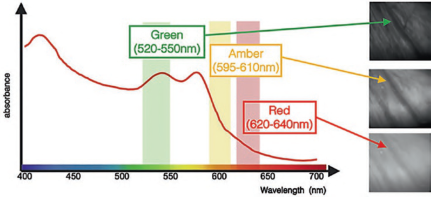
RDI utilizes long wavelength light that penetrates into deeply and visualize blood vessels and provides tissue contrast by irradiating three types of narrowband light (red, amber, and green) at different wavelengths (630 nm, 600 nm, and 540 nm, respectively) [1]. Deep and superficial blood vessels are differentiated by utilizing the differences in the absorption and reflectance of hemoglobin (. Fig. 1):
- Red (620–640 nm): Reaches deep into the mucosa at approximately 1.0 ~ 1.5 mm and displays deep blood vessels and mucosa at a constant brightness.
- Amber (595–610 nm): Reaches deep into the mucosa at approximately 1.0 ~ 1.5 mm and displays deep blood vessels.
- Green (520–550 nm): Does not reach deep into the mucosa and displays superficial capillaries.
3 Three Modes in RDI
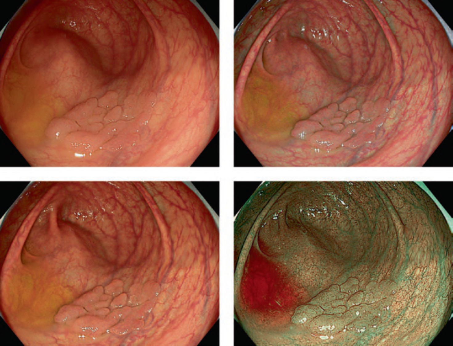
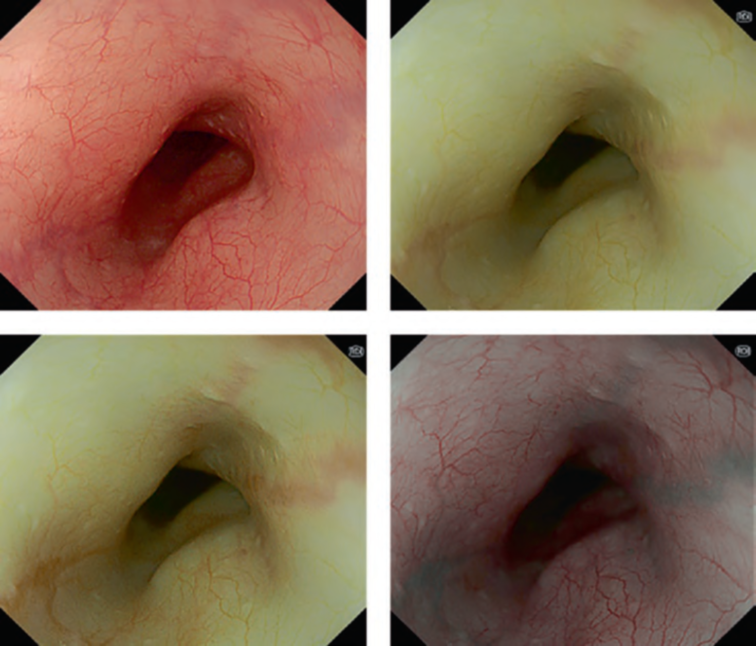
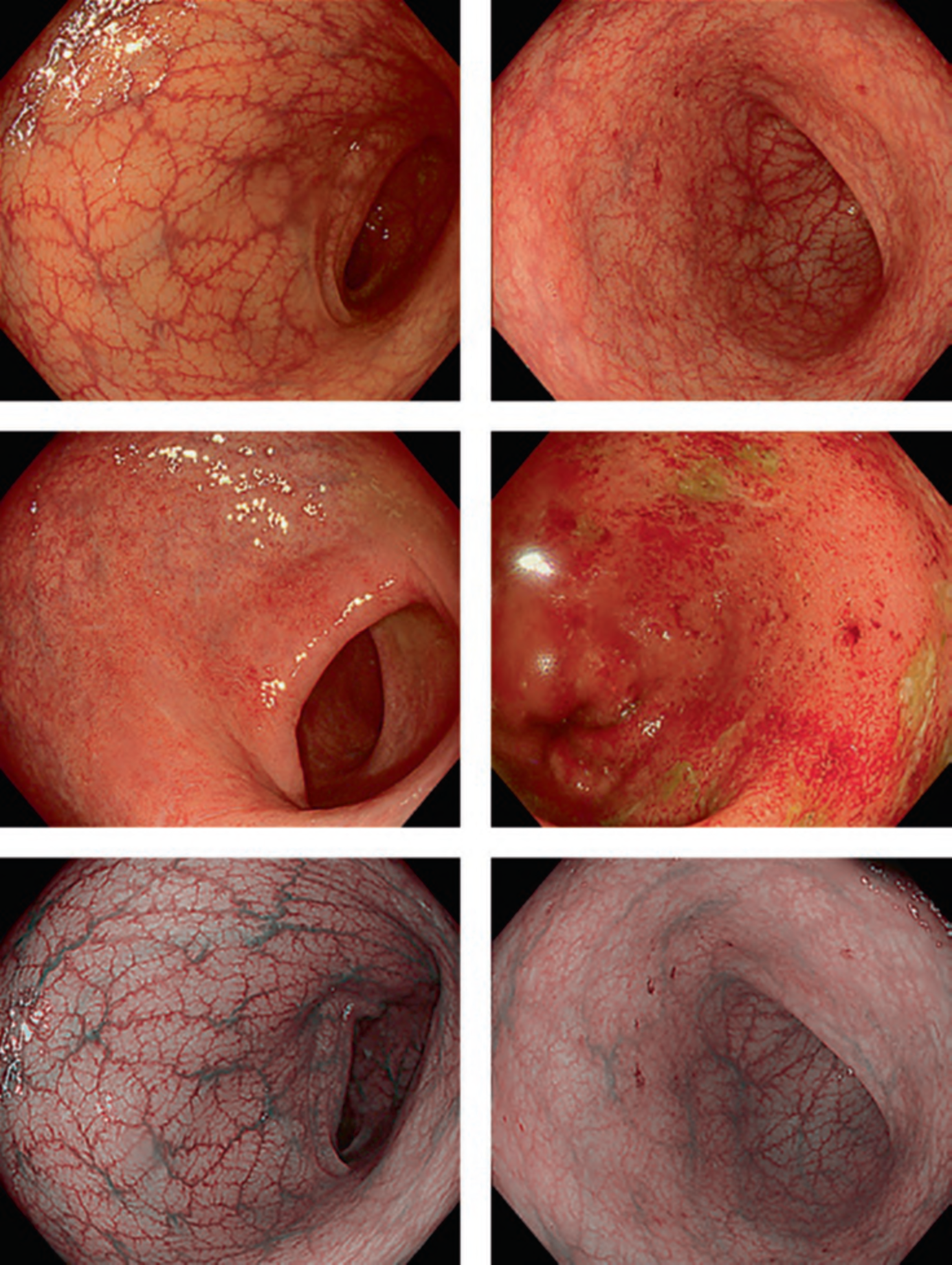
By one touch of a button on the scope, it is possible to switch from white light mode to RDI mode. RDI has three different modes, modes 1, 2, and 3 (Fig. 2). These modes can be switched in sequence by pressing a switch on the endoscope. By switching modes, it is possible to change the level of deep vessel enhancement. The features of each mode are described below.
Mode 1
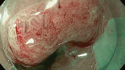
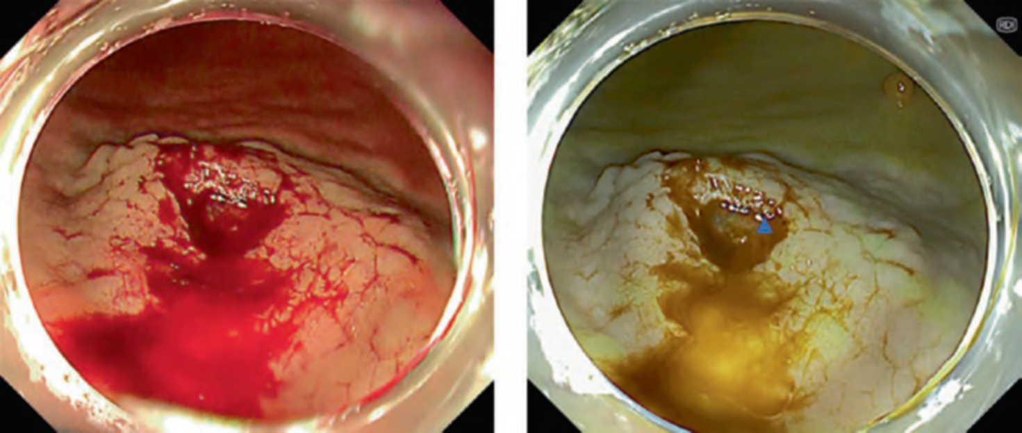
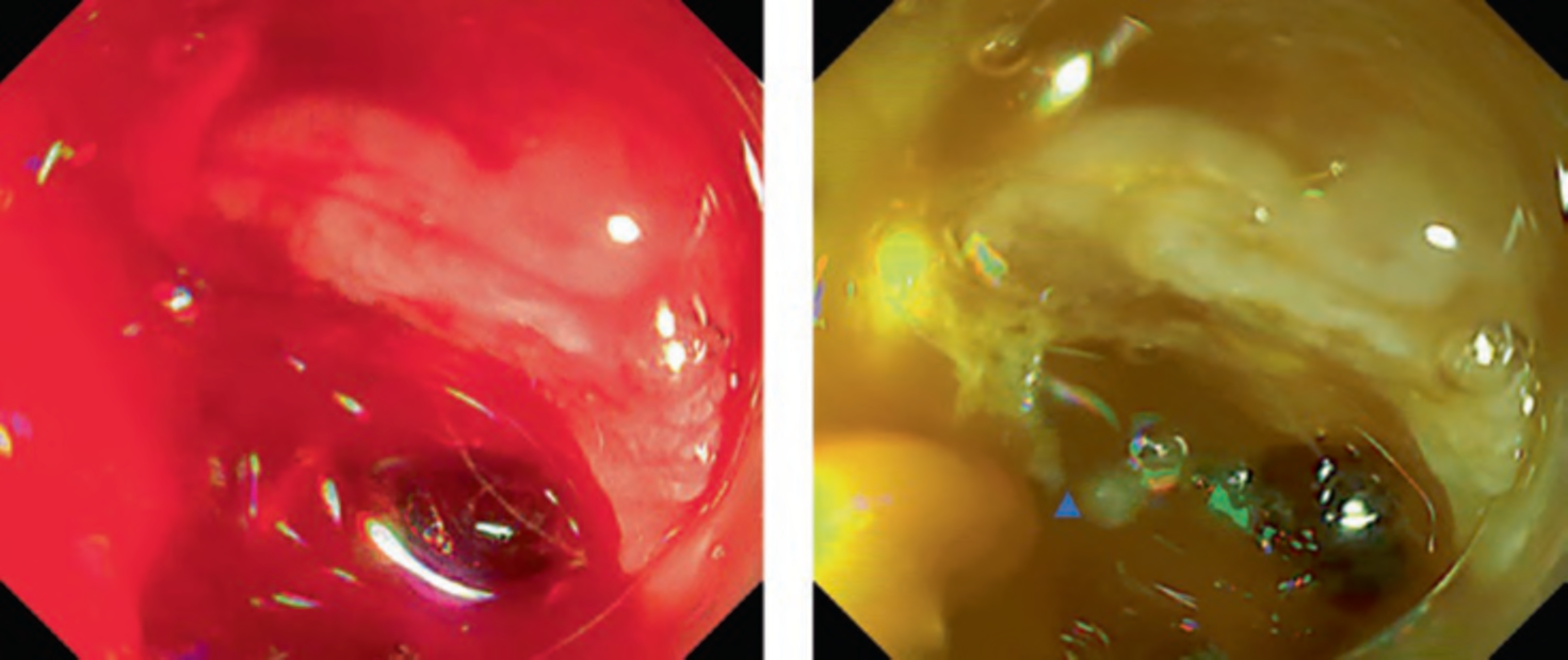
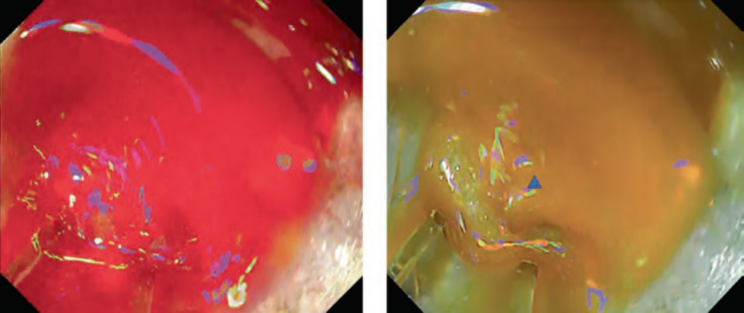
This mode is mainly aiming for identification of bleeding points. In cases of active bleeding, it is often difficult to continue the hemostasis because the pooled blood disturbs visualization. Potential usefulness of RDI in ESD by the improvement of identification of bleeding point has been reported [2–4]. A multicenter randomized controlled trial revealed significant reduction of endoscopists’ psychological stress during hemostasis even though it failed to show the shortening of hemostasis time [5]. There is also a case report of successful hemostasis for active bleeding due to peptic gastric ulcer [6] (Figs. 3, 4, and 5).
Mode 2
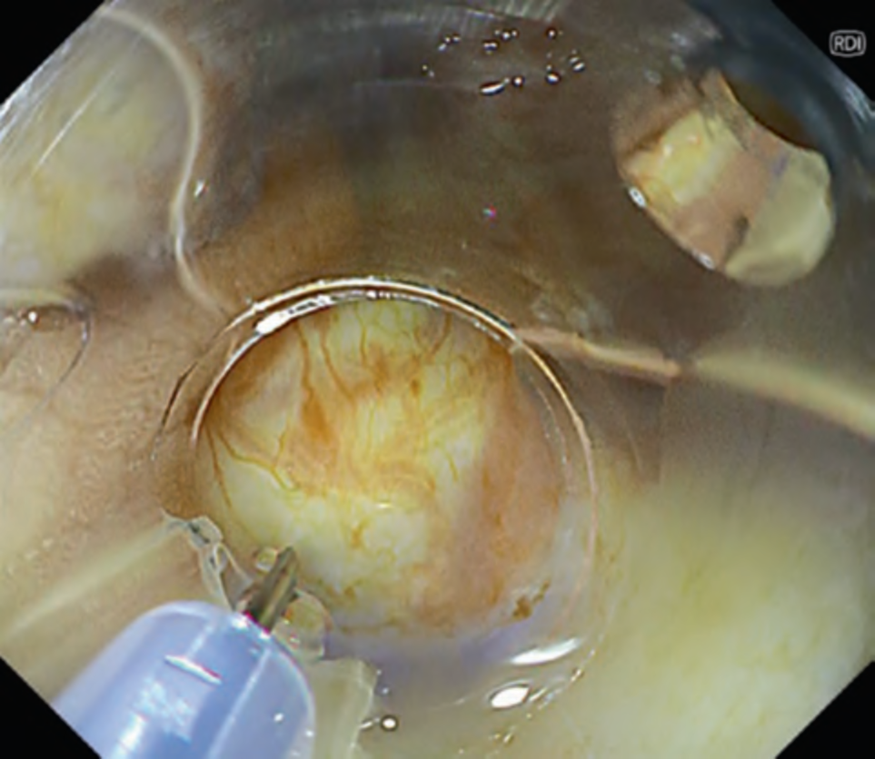
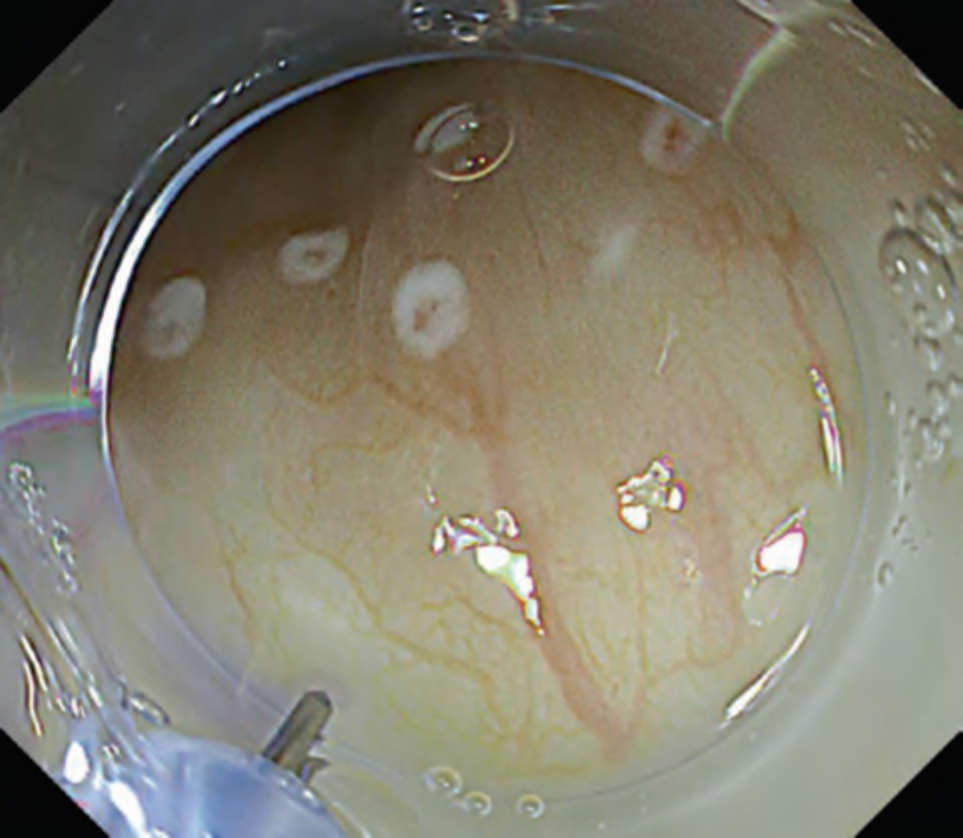
This mode is developed aiming improvement of visualization of deep blood vessels. Compared to mode 1, mode 2 enhances redness of blood vessels and improves the visibility of blood vessels in deep tissues. This is especially useful for initial submucosal injection during endoscopic submucosal dissection (ESD). Normally, when performing injection during ESD, blind injection cannot be avoided, because we cannot directly observe blood vessels below the mucosa. However, by identifying deep blood vessels under RDI mode 2 and avoiding unnecessary injury of those vessels, it prevents bleeding or the submucosal hematoma formation (. Figs. 6 and 7) [7]. Similarly, this mode is also reported to be useful for predicting the depth of esophageal varices and confirming red color signs [8].
Mode 3
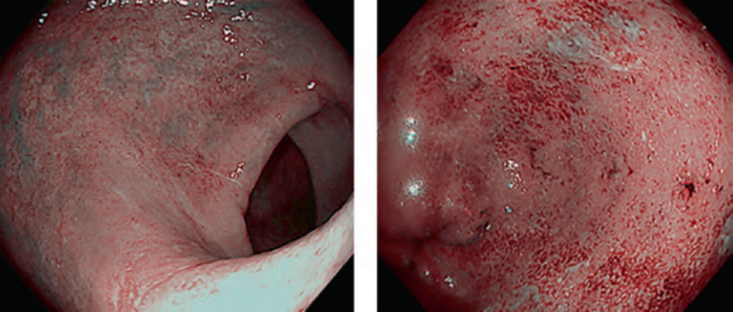
In this mode, deep blood vessels are displayed in green and superficial blood vessels in red. Images obtained by this mode are similar to those by narrowband imaging; however, bile in the digestive tract lumen does not turn red, facilitating mucosal observation in colonoscopy with poor preparation. It is also reported that this mode enables to visualize subtle inflammatory activity that is unable to find in white light imaging in patients with ulcerative colitis (. Fig. 8) [9].
4 Conclusion
In this chapter, we described the principles of RDI and its clinical applications. As mentioned above, RDI has been reported to be useful not only for initial development purpose, the endoscopic hemostasis, but also other advanced interventions and even evaluation of inflammation activity. Further application of this novel image- enhanced endoscopy technology would contribute progression of endoscopic techniques for wider and better provision for health care in the future.