

1 Introduction
The history of camera-equipped endoscopes traces back to Olympus’ prototype in 1950. An electronic endoscope capable of converting images into electrical signals and displaying them on a monitor was developed in Japan in 1986 [1]. Subsequently, models such as CV-240 (1997), CV-260, Lucera Spectrum (2006), and CV-290, Lucera Elite (2010) have evolved remarkably in terms of image quality and functionality. Notably, the recent introduction of the CV-1500, EVIS X1 (2020), marks a significant milestone after a decade and heralds further advancements (. Fig. 1). . Table 1 provides a summary of the distinctions between the previous and the latest models.
The X1 system essentially is a universal model that can be used with all types of scopes. Historically, Olympus endoscopes have been developed into two primary series due to differences in image quality demands. Frame-sequential endoscopy (Japan/UK/Hong Kong) involves a sequential image capture process employing alternating color channels, ultimately generating a full-color image, and potentially offering heightened resolution [2]. On the other hand, simultaneous endoscopy (Europe/USA) employs specialized sensors dedicated to individual color channels, allowing for the concurrent capture of images. This approach provides notable advantages, including enhanced color accuracy, diminished motion artifacts, and an overall elevation in image quality, albeit at a higher cost.
Consequently, limitations emerged in certain regions, including issues with relatively low image quality and the inability to use specific scopes such as magnifying endoscope and spiral motor power enteroscope. These problems have been resolved with the launch of the new system in 2020. In other words, it is believed that the era is changing, requiring endoscopists from all regions to be familiar with the imaging types and proficient in magnifying endoscopy diagnosis. This chapter provides a detailed description of the new features of the latest Olympus endoscopic platform, EVIS X1.
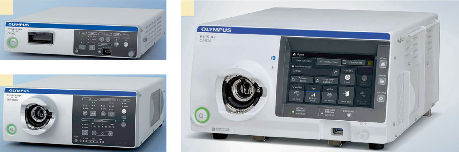
| Lucera Elite/Exera III (CV-290/CV-190) | EVIS X1 (CV-1500) | |||
| Launch | 2010 | 2020 | ||
| Light source | Xenon | LED | ||
| Compatible monitor | HD | UHD 4K | ||
| Imaging method | Frame-sequential (CV-290)/ simultaneous (CV-190) | Frame-sequential (fast spin) and simultaneous | ||
| Image sensor compatibility | Monochromatic CCD | CMOS + CCD | ||
| Image mode | WLI, NBI (second generation) AFI, IRI | WLI, NBI (third generation) AFI, RDI | ||
| Digital manipulation | None | TXI, BAI-MAC | ||
| Component | Video processor only | Video processor with light source | ||
| Weight | 9.4 kg + 18.5 kg (light source) | 19.4 kg | ||
LED light-emitting diode, HD high definition, UHD ultrahigh definition, CCD charge-coupled device, CMOS complementary metal oxide semiconductor, WLI white light imaging, NBI narrowband imaging, AFI autofluorescence imaging, IRI infrared imaging, RDI red dichromatic imaging, TXI texture and color enhancement imaging, BAI-MAC brightness adjustment imaging with maintenance of contrast
2 Features of EVIS X1
LED Light Source
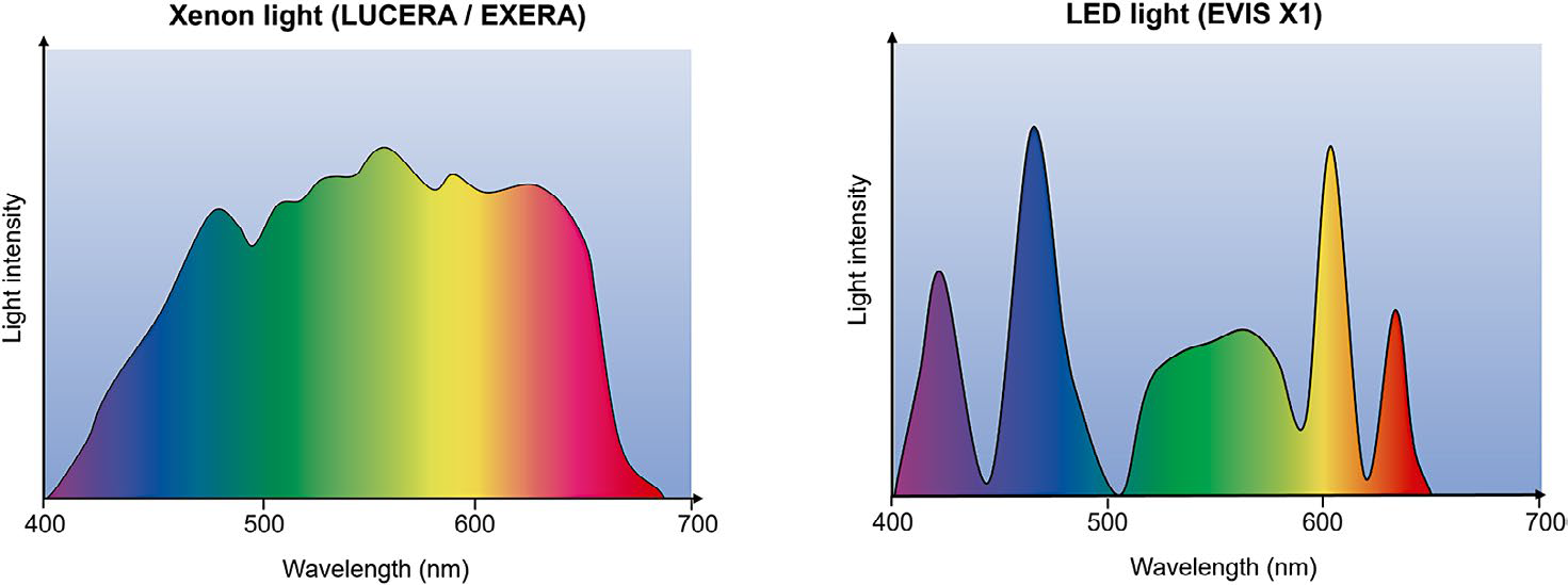
The Olympus X1 introduces a new light-emitting diode (LED) light source, fundamentally altering the emission spectrum (. Fig. 2). The LED light source integrates red, blue, yellow, violet, and a new amber color, alternately emitted and extinguished to create primary colors. These five LED light source offers a substantially extended lifespan compared to the previous xenon light source, which had a mere 500-hour lifespan. This enhancement not only prolongs the equipment’s operational life but also contributes to energy conservation. LEDs provide consistent and stable light output, ensuring uniform illumination across the field of view. This uniformity is vital for accurate visualization during endoscopic procedures, eliminating the risk of underexposed or overexposed areas. The combination of amber, red, and green enables the application of a new red dichromatic imaging (RDI) function, while blue and violet combinations facilitate narrowband imaging (NBI).
New Scopes
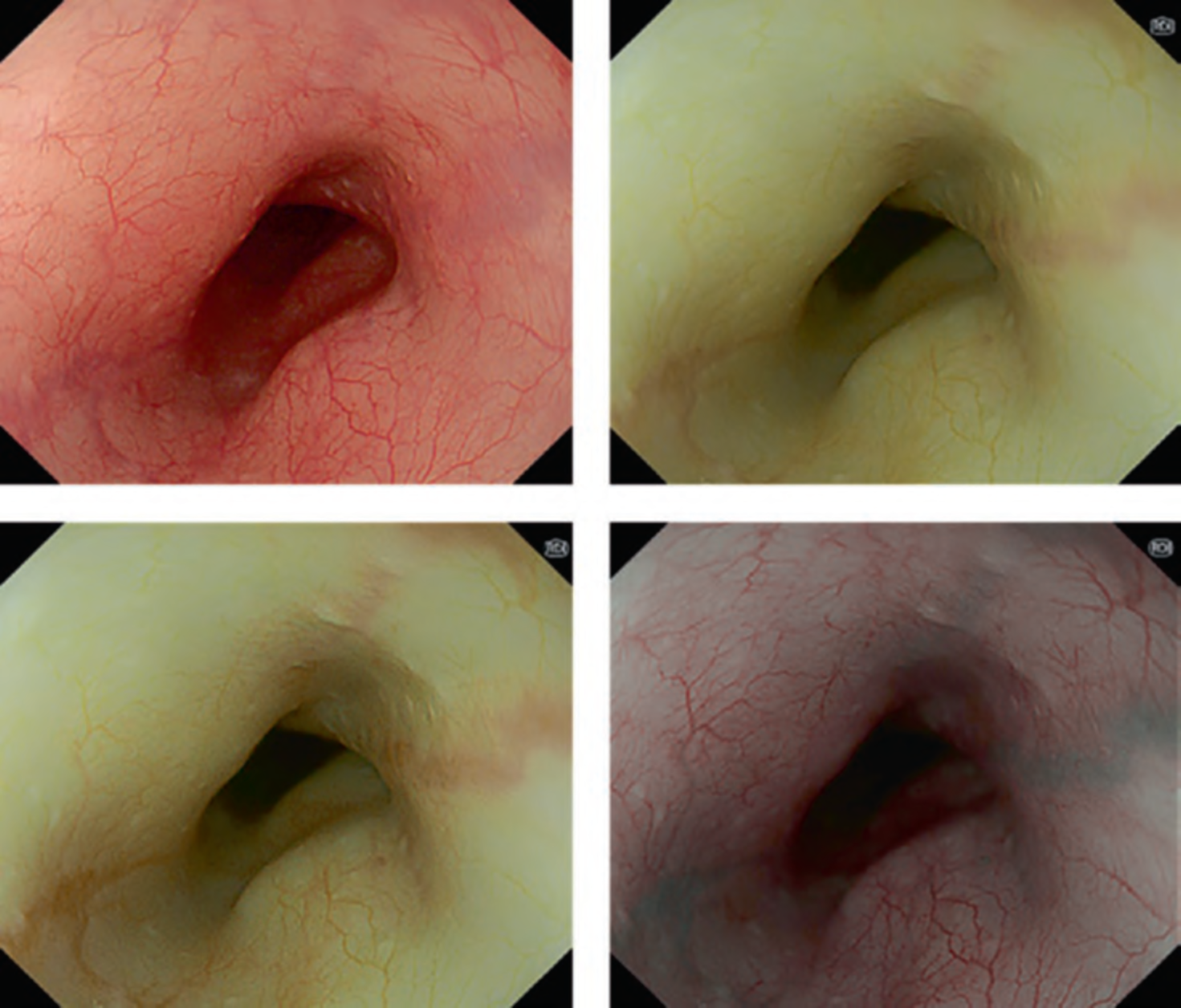
Flagship scopes for the X1 generation include GIF-EZ1500 and CF-EZ1500D models. The button-operated magnification function with dual focus from the previous GIF-HQ290 and CF-HQ290 models enabled magnification at the push of a single button, but as an optical zoom, it could only magnify up to 40×, making adequate evaluation difficult in certain cases. The new models have further enhanced functions, with magnification up to 100× and 90× for GIF and CF, respectively, and the GIF-EZ1500 allows the user to get as close as 3 mm to the subject and 1.5 mm in near-focus mode. In addition, the Expanded Depth of View (EDOF) function, which combines near and far resolution images to create a single image, provides clearer, widely focused image without demanding high-technical expertise. Moreover, the EZ-1500 scope is equipped with a newly developed color CMOS (complementary metal oxide semiconductor) sensor with high sensitivity, color reproduction, and low noise. As this scope has been widely marketed worldwide, it is predicted that magnified endoscopic diagnostics will become a standard in use. However, due to the seamless acquisition of magnified images with the new scope, there is a potential for debate regarding whether to use the classification of magnifying endoscopes or non- magnifying endoscopes, as the traditional clear distinction between non- magnified and magnified endoscopes may no longer apply.
Other models include the 1100 series, which is a non-magnifying scope with a color CCD (charge-coupled device) and is similar to the traditional Western models (Exera, 100 series), and the 1200 series, which uses a monochrome CCD and is similar to the traditional Japanese models (Lucera, 200 series) with a lever-type, high magnification function (GIF: 125×, CF: 135×, . Fig. 3). Notably, the new 1200 series incorporates high-speed rotation among its frame-sequential methods, resulting in remarkably smooth and high-resolution performance compared to the previous generations. The GIF-1200 N model is a narrow-diameter scope but uses a new CMOS sensor, which reduces noise and produces high-definition image quality similar to conventional oral endoscopes.
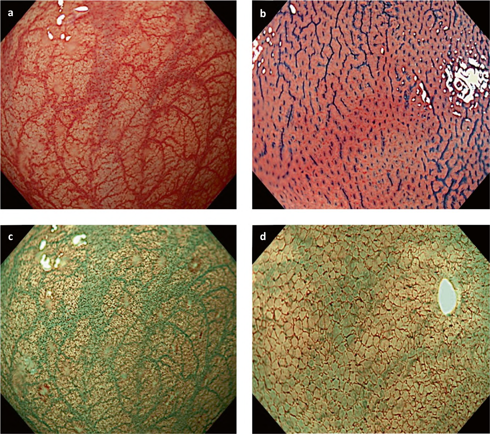
Image Enhancement
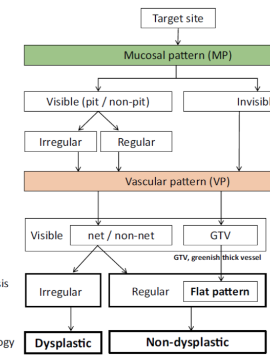
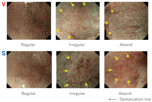
Narrowband Imaging (NBI)
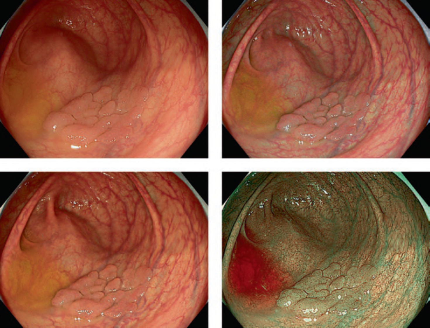
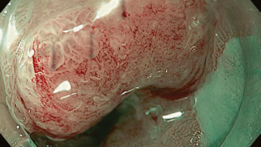
NBI involves the transformation of regular xenon white light into blue and green wavelengths. This is achieved by introducing an NBI filter between the light source and the traditional red-green-blue filter [3]. This focused light carries wavelengths of 410 nm and 540 nm and is readily absorbed by hemoglobin, rendering dense hemoglobin vessels distinctly dark. This contrast enables a detail observation of capillary network patterns vital for diagnosing colorectal issues. Furthermore, the reduction in tissue reflection and scattering elevates the visibility of surface structures.
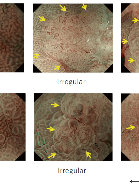
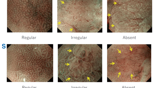
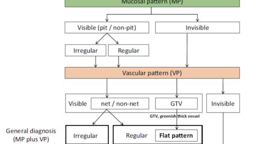
The implementation of NBI offers two primary benefits: Firstly, NBI is notably efficient and uncomplicated when compared to traditional dye-based chromoendoscopy. Given that crystal violet, a potentially carcinogenic substance, necessitates several minutes for staining, NBI enables instant observation of microstructures, devoid of potential risks. Secondly, magnifying NBI introduces a novel dimension, allowing the study of vascular network patterns alongside standard pit pattern analysis. This progress led to the creation of the Sano classification, which differentiates the type of colorectal polyps by examining their surface blood vessels [4]. As a result, the modern NBI diagnostic method combines both vascular and surface pattern information. When faced with low confidence in NBI diagnosis or when precise endoscopic prediction of invasion depth is essential for surgical decisions, the application of crystal violet staining provides supplementary clarity although there is a limited availability of the solution.
NBI, despite its advantages, has certain limitations. Particularly, its efficacy hinges on the quality of bowel preparation. In cases of inadequate bowel preparation, residual yellowish stool can be misleadingly represented as vivid red under NBI, thereby diminishing its utility. Therefore, thorough lesion washing is essential prior to undertaking detailed NBI assessment, regardless of preparation quality. Another drawback is that NBI often presents images darker than those produced by regular white light. The second-generation NBI in Lucera Elite/Exera III is brighter than the original NBI, and its effectiveness in detecting colorectal polyps has been demonstrated [5]. Due to the enhancements in LED light sources and sensors implemented in the X1, a higher level of brightness has been successfully attained. This increased brightness now provides sufficient illumination for routine practical use.
Red Dichromatic Imaging (RDI)
Red Dichromatic Imaging (RDI) is an advanced imaging technique that emphasizes red and brown colors using the newly developed amber LED light. RDI enhances the visibility of blood vessels and subtle tissue changes, contributing to improved diagnostic accuracy during endoscopic examinations [6].
Texture and Color Enhancement Imaging (TXI)
Olympus has introduced TXI in endoscopy, an innovative technique enhancing real- time visualization of tissue textures and colors. By utilizing advanced algorithms, TXI accentuates subtle patterns and color variations, aiding in the identification of tissue irregularities [7]. Using this feature can assist in the diagnosis of atrophic gastritis and is expected to improve detection rates for early gastric cancer and colorectal polyps. However, since its clinical data is not yet sufficient, future research is warranted.
Brightness Adjustment Imaging with Maintenance of Contrast (BAI-MAC)
BAI-MAC is another technology which involves digital adjustment of the brightness of endoscopic images to enhance visibility while preserving the contrast between different tissues. By iteratively fine-tuning the brightness and utilizing image enhancement algorithms, BAI-MAC helps endoscopists detect subtle abnormalities from a distance and make accurate diagnoses without compromising essential contrast information.
Application of Artificial Intelligence (AI)
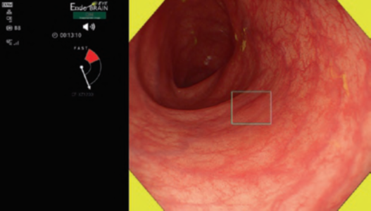
Computer-aided tools for detecting polyps, using AI and deep-learning software (CADe), have garnered significant attention in recent times, with multiple trials displaying promising results. In 2020, Olympus introduced ENDO-AID (OIP-1), a new X1 compatible CADe software that subsequently demonstrated enhanced improved adenoma detection (52.9% vs. 37.4%, p = 0.017) [8]. This innovation not only streamlines the endoscopy process but also has the potential to simplify and improve efficiency for endoscopists by minimizing the need for excessive eye movements through its user- friendly presentation of lesions.
Limitations of EVIS X1
Given the financial constraints faced by many hospitals, obtaining the latest models might not be feasible within a reasonable timeframe. Consequently, older scope models are frequently still in operation, necessitating the utilization of the previous- generation light source alongside the new X1 to maintain compatibility. Furthermore, these aging models, when projected onto expansive 4 K screens, might encounter challenges in maintaining image stability. Particularly noteworthy is the variance in image settings between these older and newer generations. While this transition can be managed through the memory function, enabling users to switch between settings, it requires manual adjustment each time the scope generation changes. Additionally, the integration of touch panel interfaces, while enhancing interactivity, introduces an added layer of intricacy when making specific adjustments.
3 Conclusions
The combination of EZ-1500 and X1 provides stress-free practice with extremely high image quality, with high levels of sharpness, brightness, and frame rate that offer perfect white light and narrowband imaging in both upper and lower gastrointestinal tract. We anticipate that the new-generation endoscopy will offer clearly defined images and seamless motion capture, contributing to a comfortable working environment for all endoscopists. The combination of a high detection rate and magnifying endoscopy is expected to yield numerous benefits for patients worldwide.