

1 Principles of TXI
Texture and color enhancement imaging (TXI) is an innovative image-enhanced endoscopy (IEE) developed by Olympus. Its core objective is to enhance the visibility of lesions by improving three vital image factors inherent to white light imaging (WLI): (1) brightness in dark areas; (2) texture, such as subtle surface changes; and (3) color, including slight color variations in the image. To achieve this, TXI applies image processing technology based on the retinex theory [1]. This theory involves decomposing images into two layers based on human visual system characteristics [2]. The first layer represents the scene’s illumination light, while the second layer corresponds to the local contrast in brightness and color.
The flowchart in Fig. 1 outlines the principles of TXI, which comprises five processes. Initially, the input image was divided into base and detailed layers in the first step. The second step corrects brightness in dark areas using the base layer. Directly adjusting brightness via light source control is challenging, as it increases the brightness throughout the entire area, causing halation in the bright regions. However, manipulating the base layer, which represents the illumination light, enables partial brightness control in specific areas. In the third and fifth steps, texture and color are enhanced, respectively. The detailed layer captures local contrast and contains information on texture and subtle surface or color changes. Texture and color can be strengthened by enhancing the detailed layers. The corrected base and enhanced detailed layers were then combined in the fourth step to generate one of the TXI outputs. Additionally, in the fifth step, the color tone was significantly enhanced, particularly for distinguishing between red and white. The output from the fifth process represents one TXI output; the other TXI output is derived from the fourth process without color enhancement, resulting in a color tone close to that of WLI. TXI offers two modes: mode 1, the output from the fifth process with color enhancement, and mode 2, the output from the fourth process without color enhancement, close to the WLI color tone [3].
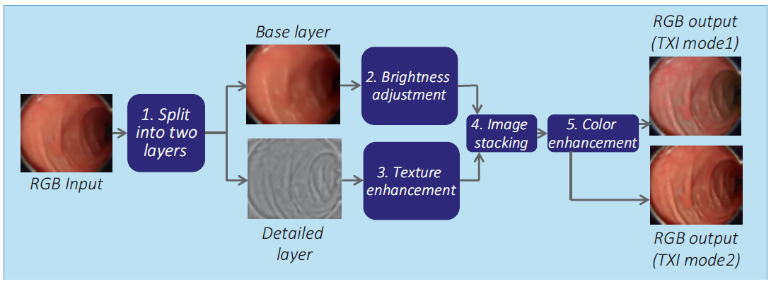
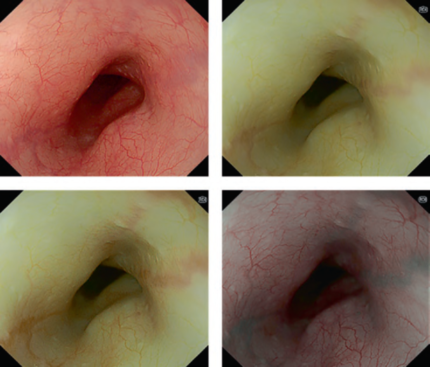
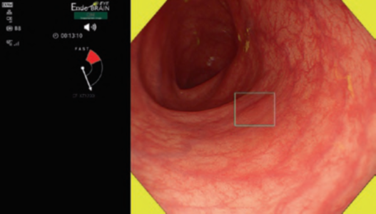
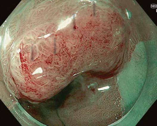
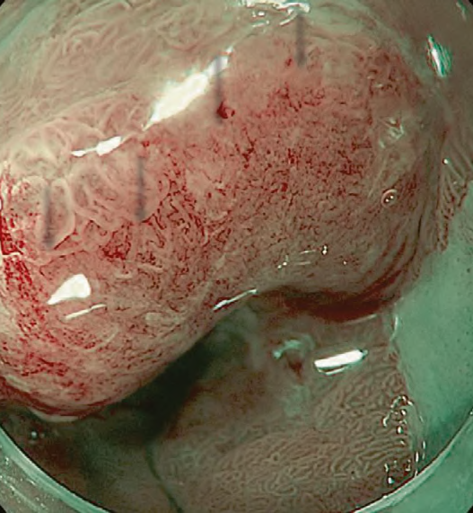
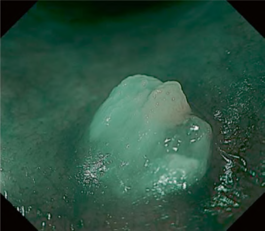
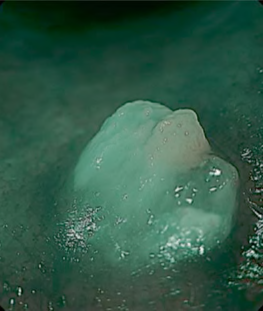
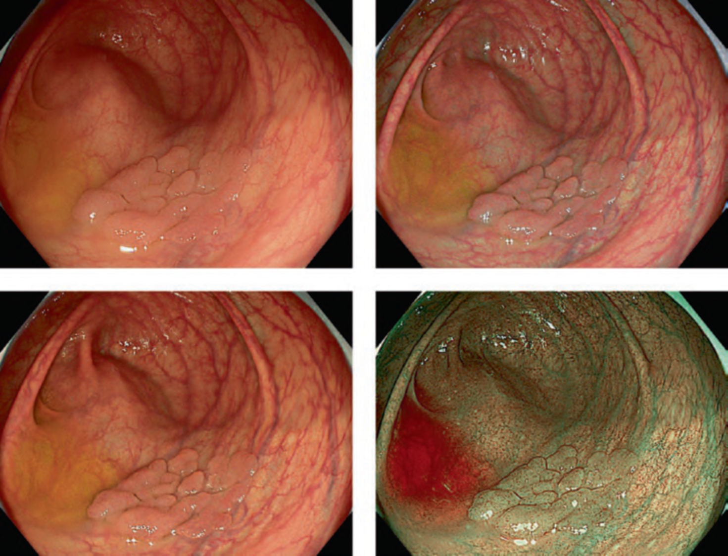
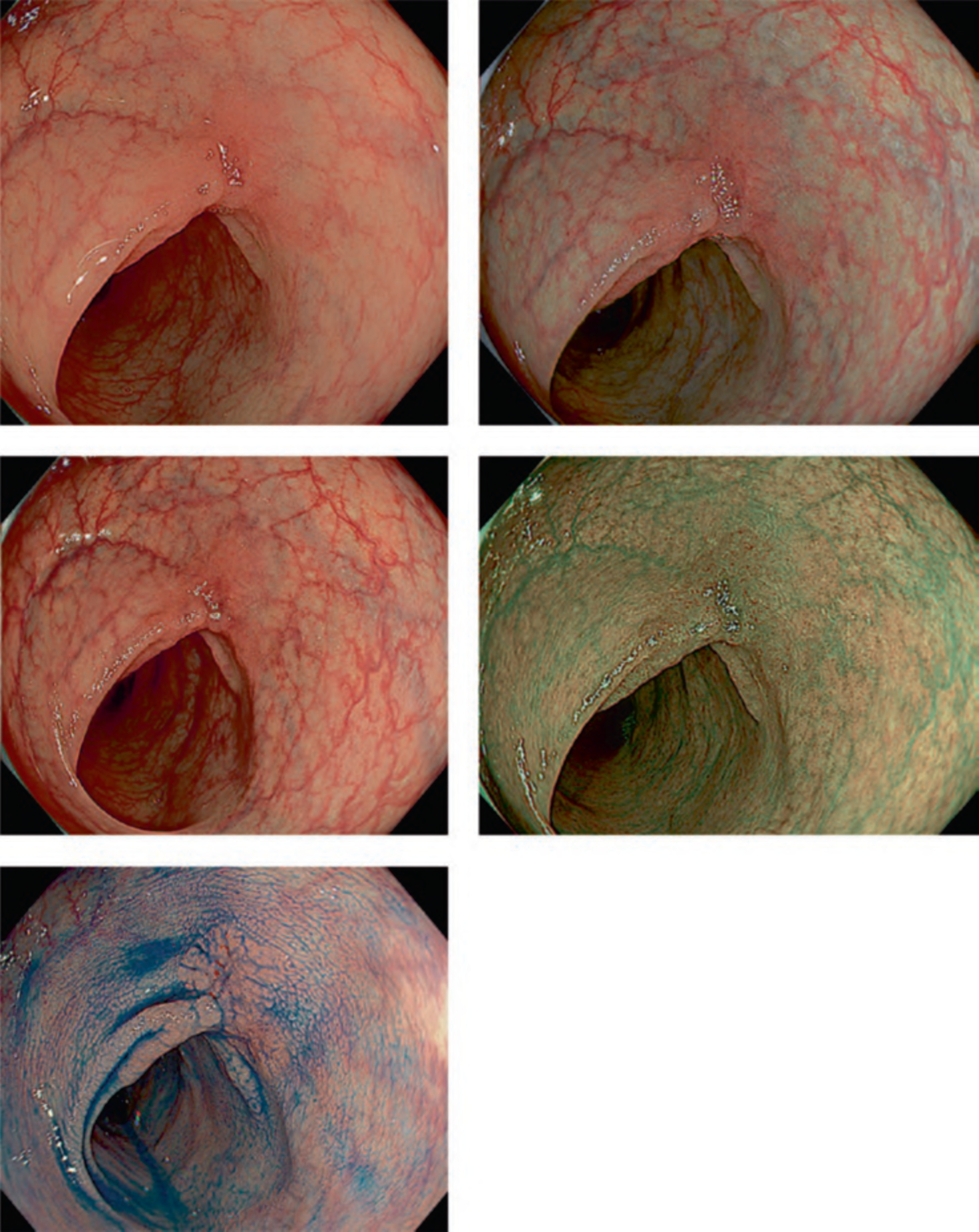
Figure 2 illustrates the features of the TXI. In TXI modes 1 and 2, gaps between the nodules were more distinctly depicted than in WLI and narrowband imaging (NBI). Furthermore, owing to the emphasis on brightness of the distal portion, the appendix orifice can be more clearly observed with TXI than with WLI. In TXI mode 1, pale areas appear even paler, and red areas appear redder, indicating a stronger contrast in color tone compared with white light. Conversely, in TXI mode 2, color tones are not emphasized, allowing for observation more similar to WLI conditions. Both TXI modes 1 and 2 offer improved visualization of vessels in the mucosa, particularly where residual liquid has accumulated. This suggests that TXI is less affected by the residual liquid than NBI. The advantage of TXI in improving visibility is also evident in non-granular laterally spreading tumors, which are often difficult to detect using WLI (Fig. 3).
2 Clinical Utility of TXI
Upper GI and Pancreaticobiliary
Studies in upper gastrointestinal endoscopy have demonstrated TXI’s efficacy in enhancing the visibility of esophageal squamous cell carcinoma [4], early gastric cancer [5], and superficial nonampullary duodenal tumors [6]. Furthermore, reports indicate that TXI enhances the effectiveness of assessing Helicobacter pylori gastritis [7] and Barrett’s esophagus [8].
In the field of pancreaticobiliary, case reports suggest that TXI improves the recognition of bile and pancreatic duct orifices [9, 10].
Colonoscopy
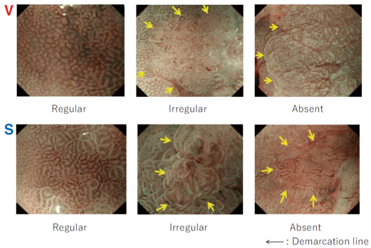
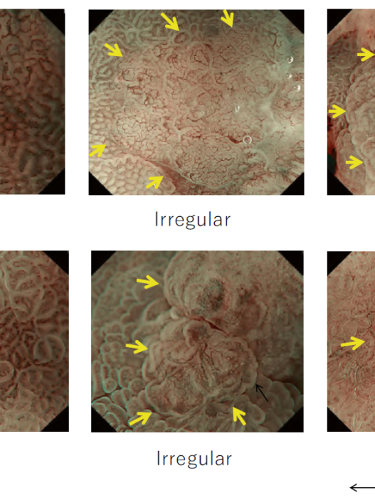
Regarding colonoscopy, research has revealed the effectiveness of TXI in providing better visualization of colorectal lesions [11] and assessing mucosal healing in patients with ulcerative colitis. Additionally [12], two studies revealed TXI’s efficacy in improving detectability of colorectal lesions. Sakamoto et al. reported that TXI improved the mean number of adenomas detected per procedure (TXI, 1.5, vs. WLI, 1.0) and adenoma detection rate (ADR) (TXI, 58.2%, vs. WLI, 46.8%) compared to WLI in a retrospective multicenter observational study [13]. Antonelli et al. conducted an international multicenter randomized trial comparing ADR between TXI and high-definition WLI groups in screening, surveillance, and diagnostic colonoscopy. They found that the TXI group had a significantly higher ADR than the WLI group (TXI, 58.9%, vs. WLI, 42.7%). Significant differences were observed for both ≤5-mm and 6–9-mm adenomas [14].
3 Future Perspective of TXI
In endoscopy, where detection and diagnosis of lesions occur on a two-dimensional monitor, TXI is an innovative IEE that facilitates the adjustment of brightness and emphasizes surface irregularities and color differences in endoscopic images. Although its effectiveness in improving the visibility of neoplastic lesions in the upper gastrointestinal tract is evident, further investigations are required to determine its potential in enhancing the detectability of neoplastic lesions. Regarding colonoscopy, TXI’s role in improving detectability of colorectal lesion has been established, and it may provide better detection of colorectal neoplasia, even in patients with poor bowel preparation. However, additional evidence is required to understand the specific conditions under which TXI excels.
The combination of TXI with chromoendoscopy or its integration with NBI has the potential to enhance endoscopic diagnostic accuracy and detectability of neoplastic lesions in the future. As evidence is accumulated, TXI is expected to significantly contribute to routine clinical practice.